Your health status is revealed through every heartbeat, and getting an ECG done is the easiest way to diagnose your heart’s condition. The electrocardiogram (ECG) functions as an essential diagnostic instrument by capturing the heart’s electrical activity, which helps healthcare providers understand critical information about heart function.
The spikes and waves visible on an ECG paper correspond to specific moments when the heart performs its function. The visual depiction of electrical signals from each heartbeat enables healthcare providers to detect different heart conditions and abnormalities.
Thus, healthcare providers and patients must understand normal versus abnormal ECG readings since these patterns can indicate cardiac issues before symptoms become severe.
The consistent use of ECG monitoring serves as a core element of preventive cardiology while enabling medical professionals to determine treatment plans for different cardiovascular diseases.
What Constitutes a Normal ECG?
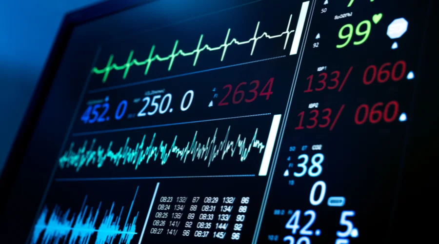
Your heart beats to its own internal rhythm similar to how a drummer maintains a consistent tempo. The display of multiple characteristic patterns in a normal ECG report demonstrates healthy cardiac activity.
Heartbeat intervals remain consistent throughout the rhythm which appears regular. Each wave component tells a specific story about the heart’s electrical activity: Atrial depolarization creates the P wave while the QRS complex illustrates ventricular depolarization.
Ventricular repolarization manifests as the T wave. The ST segment usually maintains a steady baseline as it links the QRS complex and the T wave.
A normal ECG values chart indicates that a healthy adult’s heart rate lies between 60-100 beats per minute while displaying distinct waveforms with correct amplitude and duration.
Common Signs of an Abnormal ECG
Occasionally the heart beats in an atypical rhythm. Abnormal ECG examples reveal multiple deviations from standard heart rhythm patterns.
Possible variations in an ECG can present as irregular heart rhythms known as arrhythmias or heart rates that are either too fast (tachycardia) or too slow (bradycardia) and may also show distorted waveforms.
Normal vs abnormal ECG reading assessments highlight significant differences between wave morphology and the timing and relationships of waves. Abnormal ECG results may present with enlarged or missing P waves together with widened QRS complexes and inverted T waves or ST segment deviations.
The changes observed may indicate a spectrum of cardiac conditions ranging from minor conduction problems to critical heart issues that demand urgent care. In this blog, Big Apollo Spectra, the best cardiology hospital in Patna, presents the 6 main differences between normal and abnormal ECG readings.
Quick Comparison – Normal vs Abnormal ECG
| Feature | Normal ECG | Abnormal ECG |
| Heart Rhythm | Regular and consistent | Irregular, too fast, or too slow |
| Heart Rate | Typically between 60–100 beats per minute | May be above or below the normal range |
| Electrical Activity | Normal wave patterns and intervals | Altered waveforms or unusual intervals |
| Indication | Suggests normal heart function | May indicate an underlying heart condition |
| Symptoms | Usually no cardiac symptoms | May be associated with chest pain, palpitations, dizziness, or breathlessness |
| Need for Further Testing | Often no additional tests required | May require Echo, Holter monitoring, TMT, or other cardiac evaluations |
Consult with a Cardiologist Today for Correct Diagnosis – Book Your Appointment
Normal vs Abnormal ECG – 6 Key Differences Explained
Understanding the main differences between normal and abnormal ECG readings can be informative and help patients discuss the findings with the cardiologist.
Difference 1: Heart Rate
The speed at which the heart beats provides significant information about general health conditions. The evaluation of heart rate is essential when comparing normal vs irregular ECG assessment. Normal heart rates range from 60 to 100 beats per minute but readings that fall outside this range require medical attention.
When the heart’s rate exceeds 100 bpm as tachycardia occurs, it can be related to stress or cardiac conditions while bradycardia, when the heart’s rate is below 60 bpm, suggests conduction problems or medication side effects. However, this can also be normal for highly trained athletes.
Difference 2: Rhythm
Your heartbeat serves as the body’s most dependable timekeeping mechanism. A normal ECG report displays a consistent pattern because a normal cardiac rhythm maintains regular intervals between each heartbeat.
However, abnormal heart rhythms present through irregular heartbeat intervals or occur with skipped or extra beats. Arrhythmias can manifest as chaotic patterns that suggest medical conditions such as atrial fibrillation or heart blocks that interrupt the heart’s natural pacemaker abilities.
Difference 3: P Wave
The waves on an ECG provide unique information about cardiac function. The P wave indicates atrial activation and normally shows up as a smooth rounded upward deflection before the QRS complex.
P waves that appear abnormal may display notches or enlargement and the waves can also be missing completely. The presence of these ECG waveform variations suggests atrial enlargement, ectopic atrial rhythms, or upper chamber conduction abnormalities.
Difference 4: QRS Complex
The QRS complex functions as a distinctive marker for ventricular activity. Normal and abnormal ECG interpretation commonly evaluates the specific features of the QRS complex. The QRS complex presents as normal when it maintains a duration of less than 120 milliseconds along with suitable voltage.
Abnormal QRS complexes demonstrate widening or fragmentation with atypical patterns that indicate potential ventricular conduction delays, heart muscle damage, or structural changes in the heart.
Difference 5: T Wave
Analysis of the T wave provides essential data regarding ventricular repolarization. In the majority of leads, normal T waves show an upright orientation while appearing proportional to the QRS complexes.
Abnormal T waves can take on inverted, peaked, or flattened shapes which may suggest the presence of ischemia, electrolyte problems, or other cardiac conditions.
Difference 6: ST Segment
Heart health monitoring relies on the crucial information provided by the ST segment. The ST segment typically stays flat and aligns precisely with the TP segment baseline.
What causes abnormal ECG results is changes to the ST segment which manifest as either elevation or depression. Such deviations are potential indicators of cardiac ischemia or injury as well as inflammation which necessitate immediate medical evaluation.
Common Causes of Abnormal ECGs
Your heart’s electrical patterns can be influenced by numerous different factors.
When trying to understand what are the most common ECG abnormalities, it’s crucial to evaluate multiple factors, such as coronary artery disease together with electrolyte imbalances and medications as well as structural heart issues that commonly cause these ECG abnormalities.
Other contributors that impair cardiac function and cause ECG abnormalities include:
- hypertension,
- genetic conditions,
- and systemic diseases affecting cardiac function.
It’s important to note that patterns on ECGs can also be affected by environmental factors, such as extreme temperatures along with dehydration and physical stress. Healthcare providers can select proper diagnostic and treatment approaches by understanding the causes of ECG abnormalities.
Can You Have Heart Problems Even With a Normal ECG?
It is important to note that even if your ECG is quite normal, it doesn’t necessarily rule out the possibility of heart disease. Some quite serious ailments in the heart such as coronary artery disease, problems with heart valves, or even certain types of arrhythmias could be present without being indicated by a resting ECG.
Often, the heart might indicate functionally abnormal signs only during stress or physical activity, and this explains why doctors may go for stress tests combined with ECGs.
On the other hand, ailments that affect a heart’s structure rather than its electrical functions, for instance cardiomyopathies or valve diseases, are also barely detectable by standard ECGs.
If you exhibit heart disease symptoms such as chest pain, breathlessness, or palpitations even after a normal ECG result, your healthcare provider may consider more tests such as echocardiograms, stress tests, or cardiac CT scans.

Interpreting an ECG: The Role of Healthcare Professionals
To interpret an ECG correctly, you need the expertise and experience of medical professionals.
Thus, if you’re worrying about abnormal ECG findings, know that while learning about the above parameters is useful for informational purposes, you shouldn’t completely rely on personal interpretations.
To determine whether an ECG report displays normal results, it’s paramount to understand the different elements of cardiac electrical activity, and only a healthcare professional has that level of expertise. Thus, qualified healthcare professionals must perform professional interpretations to achieve accurate diagnoses.
The best cardiologist in Patna at the Big Apollo Spectra Hospital can help with the proper ECG diagnosis and interpretation with an extensive understanding of cardiac physiology and pathology.
Healthcare providers evaluate clinical context and patient’s history alongside further diagnostic tests when abnormalities appear to create appropriate management strategies.
What Happens If Your ECG Report Is Abnormal?
An abnormal ECG reading doesn’t necessarily mean you have a critical heart issue; it indicates that further tests are required. Your cardiologist will consider your symptoms, past health, and risk factors in conjunction with your ECG results to understand what is actually going on.
You may be referred for additional investigations like echocardiograms, treadmill tests, blood analysis, or cardiac imaging, based on the particular abnormalities detected.
Several ECG changes necessitate starting treatment without delay, whereas some are just observed for changes over time. The main thing is not to worry; rather, comply with your heart specialist’s instructions for more tests and therapy.
Identifying problems early via abnormal ECG results generally improves the prognosis and makes the evolution of advanced complications unlikely.
Schedule Your Cardiology Consultation Now →
FAQs:
1. How can you tell if an ECG is normal or abnormal?
A normal ECG shows a regular heart rate of 60-100 bpm, consistent rhythm with evenly spaced beats, smooth P waves, QRS complexes under 120 milliseconds, upright T waves, and a flat ST segment baseline; abnormal ECGs deviate from these patterns with irregular rhythms, rate changes, or wave shape distortions.
2. Is an abnormal ECG serious?
Not necessarily. Abnormal ECGs range from minor findings requiring only monitoring to serious conditions demanding immediate treatment; your doctor’s interpretation based on your symptoms and medical history determines the actual significance.
3. Can stress cause abnormal ECG readings?
Yes, stress can temporarily cause ECG changes like increased heart rate or T wave abnormalities; however, persistent abnormalities suggest underlying cardiac conditions rather than stress alone.
4. Can ECG detect blockage?
An ECG can show signs of ischemia (reduced blood flow) that suggest blockage, particularly during a stress test, but it cannot directly visualize blocked arteries, coronary angiography or CT angiography is needed for definitive blockage detection.
5. How long does an ECG take?
A standard 12-lead ECG typically takes 5-10 minutes from preparation to completion, with the actual recording lasting only about 10 seconds.
Get a Clear Picture of Your Heart’s Health – Contact Big Apollo Spectra
Your heart deserves expert attention and care. Performing regular ECG monitoring provides essential support for early detection and treatment of heart conditions. Get professional medical evaluation immediately when you encounter concerning symptoms or abnormal ECG results.
Big Apollo Spectra’s Cardiology department features expert professionals who deliver complete heart care using advanced diagnostics and individualized treatment approaches for optimal cardiac health. Early recognition of cardiac issues followed by prompt treatment tends to produce improved results in cardiac health management.